Diagram Of Shoulder Ligaments / Shoulder And Axilla Amboss / Printable shoulder muscles diagrams to help you study the muscles structure in human's shoulder.we have five muscle diagrams of the shoulder.
Diagram Of Shoulder Ligaments / Shoulder And Axilla Amboss / Printable shoulder muscles diagrams to help you study the muscles structure in human's shoulder.we have five muscle diagrams of the shoulder.. Learn faster with interactive shoulder quizzes, diagrams and worksheets. Each of these muscles has its origin on the scapula and inserts around the head of the. A ring of cartilage known as the labrum surrounds the glenoid fossa to extend the size of the socket while maintaining flexibility. The primary function of the shoulder girdle is to give strength and range of motion to the arm. One or more ligaments provide stability to a joint during rest and movement.
17 photos of the diagram of shoulder muscles and tendons. Diagram of the human shoulder joint. Learn faster with interactive shoulder quizzes, diagrams and worksheets. Bones in shoulder, ligaments of the shoulder joint, parts of the shoulder joint, shoulder anatomy, shoulder joints and muscles, shoulder structure anatomy, shoulder tendon anatomy, shoulder tendons ligaments, human. In the shoulder joint, the ligaments play a key role in stabilising the bony structures.
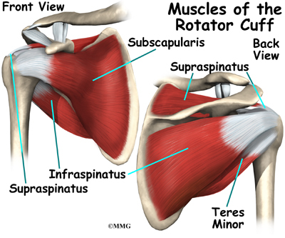
Shoulder Injuries Shoulder Pain Information Sinew Therapeutics from sinewtherapeutics.com Neck muscle anatomy mri 12 photos of the neck muscle anatomy mri neck muscle anatomy images, neck muscle anatomy pictures, neck muscle anatomy posterior, neck muscle anatomy ultrasound, neck muscles anatomy radiology, human muscles, neck muscle anatomy images, neck muscle anatomy pictures, neck muscle anatomy. A torn ligament is one such injury that an individual tends to have. The supraspinatus, infraspinatus, teres minor and subscapularis muscles are known as the shoulder rotator muscles. Diagram of the shoulder, including the location of the rotator cuff. They include the coracoclavicular ligaments, the coracoacromial ligaments, the superior transverse scapular ligament, the coracohumeral ligament, the acromioclavicular ligament, and the glenohumeral ligaments. When the shoulder dislocates, the ligaments of the shoulder capsule can be torn. It is due to the lack of a take an example of your elbow joint. The coracohumeral, glenohumeral ligaments and the tendons of the supraspinatus and subscapularis muscles all serve …
Supraspinatus, infraspinatus, subscapularis, and teres minor.
To ensure proper range of motion, the shoulder joint is supported by the shoulder ligaments, shoulder tendons and shoulder muscles. Ligaments are soft tissue structures that connect bones to bones. Diagram demonstrating the ligaments involved during the pronation and supination of the elbow. The collection of muscles and tendons in the shoulder is known as the rotator cuff. The shoulder girdle includes three bones—the scapula, clavicle and humerus. Diagram of the shoulder, including the location of the rotator cuff. Although most people think of the shoulder as a single joint between the upper arm bone (humerus) and the torso, the shoulder actually has several smaller joints outside the arm bone's socket. In the shoulder joint, the ligaments play a key role in stabilising the bony structures. When the shoulder dislocates, the ligaments of the shoulder capsule can be torn. Several ligaments make up parts of the joint capsule, and these ligaments are important in keeping the shoulder joint in proper position. Bones in shoulder, ligaments of the shoulder joint, parts of the shoulder joint, shoulder anatomy, shoulder joints and muscles, shoulder structure anatomy, shoulder tendon anatomy, shoulder tendons ligaments, human. Diagram of the human shoulder joint. The shoulder joint is formed where the humerus (upper arm bone) fits into the scapula (shoulder blade), like a ball and socket.
The tear in the ligament makes the shoulder unstable and it becomes difficult to carry out activities of daily living or recreational activities. Although most people think of the shoulder as a single joint between the upper arm bone (humerus) and the torso, the shoulder actually has several smaller joints outside the arm bone's socket. Supraspinatus, infraspinatus, subscapularis, and teres minor. A ligament in the shoulder can rupture due to various reasons. Printable shoulder muscles diagrams to help you study the muscles structure in human's shoulder.we have five muscle diagrams of the shoulder.
Shoulder Anatomy Eorthopod Com from eorthopod.com Diagram of the human shoulder joint. The glenoid fossa forms a very shallow socket, so the muscles, ligaments, and cartilage of the shoulder joint reinforce its structure and help to prevent dislocations. Although most people think of the shoulder as a single joint between the upper arm bone (humerus) and the torso, the shoulder actually has several smaller joints outside the arm bone's socket. The shoulder has about eight muscles that attach to the scapula, humerus, and clavicle. Tendons of these muscles come together to form a covering around the head of the upper arm bone (humerus) and top of the shoulder. A ring of cartilage known as the labrum surrounds the glenoid fossa to extend the size of the socket while maintaining flexibility. Related posts of diagram of shoulder muscles and tendons neck muscle anatomy mri. Several ligaments make up parts of the joint capsule, and these ligaments are important in keeping the shoulder joint in proper position.
17 photos of the diagram of shoulder muscles and tendons.
These muscles form the outer shape of the shoulder and underarm. One or more ligaments provide stability to a joint during rest and movement. Printable shoulder muscles diagrams to help you study the muscles structure in human's shoulder.we have five muscle diagrams of the shoulder. The shoulder is not a single joint, but a complex arrangement of bones, ligaments, muscles, and tendons that is better called the shoulder girdle. Related posts of diagram of shoulder muscles and tendons neck muscle anatomy mri. Diagram of shoulder ligaments : The fixture included an additional aluminum plate (c) which was connected and moved with the instron actuator. The supraspinatus, infraspinatus, teres minor and subscapularis muscles are known as the shoulder rotator muscles. The rotator cuff consists of four muscles: Their tendons blend with each … The joints of the shoulder that are primarily responsible for movement are held together by several strong ligaments. The glenoid fossa forms a very shallow socket, so the muscles, ligaments, and cartilage of the shoulder joint reinforce its structure and help to prevent dislocations. The shoulder has about eight muscles that attach to the scapula, humerus, and clavicle.
One or more ligaments provide stability to a joint during rest and movement. Related online courses on physioplus. The shoulder joint permits a fuller range of motion than any other joint, allowing the arm to raise, lower, extend and rotate a full 360 degrees. 2 ligaments (trapezoid& conoid ligaments) attach the clavicle coracoid process of scapula these tiny ligaments (w/ acominoclavicular joint) keep scapula attached to clavicle. Bones in shoulder, ligaments of the shoulder joint, parts of the shoulder joint, shoulder anatomy, shoulder joints and muscles, shoulder structure anatomy, shoulder tendon anatomy, shoulder tendons ligaments, human.
Shoulder Ligaments And Tendons Diagram Quizlet from o.quizlet.com The bicep has two shoulder tendons: This diagram with labels depicts and explains the details of ligaments of the shoulder joint. In human anatomy, the shoulder joint comprises the part of the body where the humerus attaches to the scapula.1 the shoulder is the group of structures. Printable shoulder muscles diagrams to help you study the muscles structure in human's shoulder.we have five muscle diagrams of the shoulder. Diagram demonstrating the ligaments involved during the pronation and supination of the elbow. In the shoulder, the joint capsule is formed by a group of ligaments that connect the humerus to the glenoid. The left shoulder and acromioclavicular joints, and the proper ligaments of the scapula. License image the joint cavity is surrounded by a loose fitting fibrous articular capsule.
A torn ligament is one such injury that an individual tends to have.
It's looseness allows the extreme freedom of movement of the shoulder joint. The tear in the ligament makes the shoulder unstable and it becomes difficult to carry out activities of daily living or recreational activities. The left shoulder and acromioclavicular joints, and the proper ligaments of the scapula. Supraspinatus, infraspinatus, subscapularis, and teres minor. The rotator cuff muscles are important stabilizers and movers of the shoulder joint. Fall on one point of shoulder and can rupture these ligaments with dislocation of ac joint. Diagram of the human shoulder joint. Printable shoulder muscles diagrams to help you study the muscles structure in human's shoulder.we have five muscle diagrams of the shoulder. A ligament in the shoulder can rupture due to various reasons. The supraspinatus, infraspinatus, teres minor and subscapularis muscles are known as the shoulder rotator muscles. The collection of muscles and tendons in the shoulder is known as the rotator cuff. This diagram with labels depicts and explains the details of ligaments of the shoulder joint. Tendons of these muscles come together to form a covering around the head of the upper arm bone (humerus) and top of the shoulder.
Muscles and ligaments of the shoulder poster diagram of shoulder. The torn or strained ligament is really millions of tears of these strands which are molecules of collagen.


0 Komentar